Caudal epidural injections are a well established treatment for acute sciatica, delivering anti inflammatory medication close to the affected nerve root to help settle pain and allow rehabilitation to progress.
Traditionally, these injections have been performed under fluoroscopy, or X-ray guidance, to confirm accurate needle placement. But a growing body of evidence shows that ultrasound guidance can achieve the same accuracy and outcomes, with some important advantages for patients and for the wider healthcare system.
What is a caudal epidural injection?
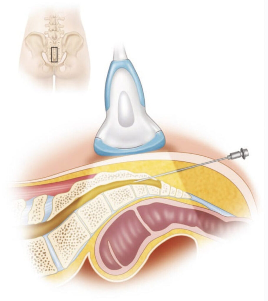
A caudal epidural injection delivers medication into the epidural space through the sacral hiatus, a small natural opening near the base of the spine. It is commonly used for sciatica caused by nerve root irritation or inflammation, aiming to reduce pain enough to allow active rehabilitation to progress.
Why has fluoroscopy traditionally been used?
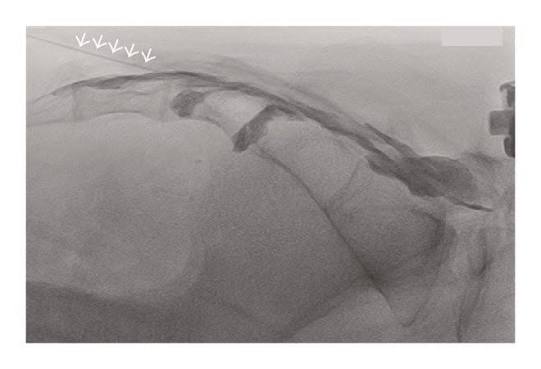
Fluoroscopy allows a clinician to watch the needle and the spread of contrast dye in real time, which has made it the traditional gold standard for confirming accurate placement.
However, it comes with real practical costs. It requires access to a hospital theatre or radiology suite, exposes the patient to radiation, and adds to waiting times and NHS capacity pressures.
What does the evidence say about ultrasound guidance?

A number of studies have now examined whether ultrasound can match fluoroscopy for accuracy and outcomes:
- Chen (2004): Ultrasound guided needle placement was correct in 100% of cases, confirmed against fluoroscopy contrast.
- Park (2015): A randomised controlled trial found ultrasound guidance was as effective as fluoroscopy for pain and functional outcomes.
- Klunklin (2022): In 110 patients with sciatica, 83% had more than a 50% reduction in pain at 24 weeks, with disability scores almost halved.
- Bernadotto (2026): Ultrasound-guided spinal injections (caudal epidural, facet joint and nerve root injections) can be safely delivered in an outpatient clinic setting.
- Systematic reviews report success rates consistently between 97 and 100% across different clinical settings.
Taken together, this evidence suggests that in appropriately selected patients, ultrasound guidance is a genuinely reliable alternative to fluoroscopy for caudal epidural injections.
Why this matters for the NHS
Fluoroscopy requires hospital theatre or radiology suite access, which adds cost and lengthens waiting times.
Ultrasound equipment is portable, involves no radiation exposure, and can be used safely in outpatient and community clinic settings, without the need for a specialist imaging suite.
This matters beyond convenience. It aligns directly with the NHS Long Term Plan’s aim of moving appropriate care out of hospitals and closer to patients, improving access and efficiency without compromising safety or accuracy.
Is ultrasound guidance right for every patient?
Not necessarily. Patient selection still matters, and ultrasound guidance relies on appropriate training, experience, and clear anatomical landmarks.
As with any injection technique, the decision should be based on the individual patient’s diagnosis, anatomy, and clinical presentation, not simply on which equipment is available.
Bottom line
Ultrasound guided caudal epidural injections offer accuracy comparable to fluoroscopy for the treatment of acute sciatica, without the need for radiation or hospital based imaging.
For appropriately selected patients, this approach can mean faster access to effective treatment, delivered closer to home, in line with the direction the NHS is already moving.